- stimulation of receptors in skin and muscle is transferred to spinal sensory nerves (found in the dorsal ganglion), which carry the stimlation through the sensory tracts of the spinal cord
- some take part in spinal reflexes, these pass into the ventral horn and synapse with motor neurons there.
- most continue on the ascending tracts and cross-over in the spinal cord or the medulla oblongata and enter cerebral centers on the opposite sides.
- stimulation from the touch and heat receptors of the skin enters the dorsal horn, then cross-over in the spine before continuing along the lateral ascending tracts to the thalamus.
- sensory information from the deeper layers of the skin and the muscles enters the dorsal ascending tracts to the medulla oblongata, there the neurons synapse, then cross-over and travel along the opposite side to the thalamus
- information is processed and grouped in the thalamus, then sent on to the appropriate parts of the cerebrum.
 |
| http://faculty.southwest.tn.edu/rburkett/A&P1_nervous_syst_organization.htm |
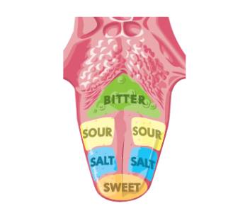 |
| http://chargedmagazine.org/2012/09/taste-buds/ |
 |
| http://www.allthingsherbal.biz/2011/10/aromas-and-the-sense-of-smell/ |
- stimulation of the cerebral olfactory nerves goes directly to the cerebrum without passing through the thalamus
Sight receptors are the eyes! They contain many light receptors, but the eye is more complex that simply being a mass of light receptors.
The eyeball is surrounded by 3 layers:
- the outer layer is protective, the sclera (ínhártya) is is the white layer, which becomes the clear cornea (szarúhártya) at the front of the eye. Light passes through the cornea to the inside of the eye. The cornea is constantly moistened by the blinking of the eyelids.
- the middle layer contains many blood capillaries, providing the eye with necessary oxygen and nutrients, it also absorbs excess light to prevent damage. It is called the choroid (érhártya). At the front it forms the ciliary body, which to which the muscles controlling the lens attach. The iris (szivárványhártya) is a continuation of the ciliary body and the hole in the middle, through which light passes into the eye is called the pupil. The colour of the iris depends on the number of pigments it contains. If someone's eyes are black or brown, then the iris contains many pigments, while people with green or blue eyes contain fewer pigments.
- the inner layer is where the receptors are found. It is called the retina (ideghártya) and it contains two kinds of light receptors, rods and cones.
 |
| http://libot-libot.blogspot.hu/2011/05/cataract-is-clouding-of-lens-of-eye.html |
 |
| http://lpatersonbiotask3.wikispaces.com/Describe+the+anatomy+and+function+of+the+human+eye... |
- light passes through several different media before it reaches the receptors
- First the light passes through the cornea
- Then it passes through space between the cornea and the lens, which contains the aqueous humour (csarknovíz), which is watery in consistency
- The size of the pupil decreases when there is lots of light, due to the constriction of the iris, and increases in low light, due to relaxation of the iris.
- Light then passes through the lens (just behind the iris). The lens is flexible and can be flattened or rounded to focus the light on the fovea (sárgafolt) at the back of the retina. This is the point in the eye of truly focused vision
- When the eye is relaxed then focus is on distant objects and the muscles holding the lens are constricted, so the lens is flattened, the opposite occurs when focusing on close objects, the muscles relaxes and the lens becomes rounded. With age, the flexiblilty of the lens decreases, so most elderly people have good distant vision, but difficulty focusing close objects. Focal problems can and often do occur earlier in life and can be corrected with proper optical lenses. If someone is far-sighted the focal point is behind the retina, while if someone is near-sighted the focla point is in front of the retina.
- the chamber behind the lens is filled with the vitreous humour, which is a jelly-like substance that helps maintain the eye's shape.
- when light reaches the retina, it activates the receptors, the rods (pálcikák) and cones (csapok), which got their names from their unique shapes.
- Rods are long and cylindrical. They work well in low light and produce black and white images.
- Cones are smaller and conical. They function well in bright light and allow us to see in colour.
- When photons are "caught" by the receptors, their energy is transformed into an electrical impulse, which is transfered down the receptor cell's membrane to the synapse. The stimulation is then carried by sensory neurons in the optic nerve to the thalamus. From here, via synapses, the stimulation is sent to the visual areas in the occipital lobes.
Rods and cones are organized as shown below. Where the optic nerve enters the back of the eye, there is a point on the retina where there are no rods or cones. This is the blind spot.
 |
| http://www.biochemj.org/csb/010/csb010_fig071.htm |
 |
| http://tiffanybiology.blogspot.hu/2011/05/rods-and-cones.html |
 |
| http://abcarcade.com/blindspot-test.html |
Sound and balance receptors are found in the ears.
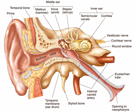 |
| http://www.biographixmedia.com/human/ear-anatomy.html |
- The outer ear's shape is designed to bring sounds into the auditory canal to the ear drum (also called tympanic membrane).
- The ear drum is the beginning of the middle ear. In the space behind the ear drum, the smallest bones in the body called ossicles, the hammer, anvil and stirrup, can be found. The hammer moves when a sound vibration causes the ear drum to vibrate. The movement of the hammer moves the anvil, which in turn, moves the stirrup. The bottom of the stirrup hits the oval window (not labelled in upper diagram) of the inner ear.
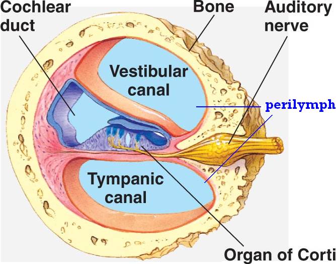
- The inner ear begins with the oval window and beyond it, the cochlea, which is filled with fluid called perilymph. When the stirrup hits the oval window, this begins a wave in the cochlear fluid. The round window, at the other end, gives the fluid somewhere to go.
- The basilar membrane is a rigid surface that extends the length of the cochlea. When the stirrup moves in and out, it pushes and pulls on the basilar membrane that is right below the oval window. This force starts a wave along the surface of the membrane. The basilar membrane has a peculiar structure. It is made of about 25 000 reed-like fibers that extend the width of the cochlea. Near the oval window the fibers are short and stiff. Toward the other end the fibers are longer and more flexible. Thus the fibers have different resonant frequencies. A specific wave frequency will resonate perfectly with the fibers at a certain point, causing them to vibrate rapidly. When the wave reaches the fibers with the same resonant frequency, the wave's energy is suddenly released. Because of the increasing length and decreasing rigidity of the fibers, higher-frequency waves vibrate close to the oval window and lower frequency waves vibrate at the other end of the membrane.
- The basilar membrane is a rigid surface that extends the length of the cochlea. When the stirrup moves in and out, it pushes and pulls on the basilar membrane that is right below the oval window. This force starts a wave along the surface of the membrane. The basilar membrane has a peculiar structure. It is made of about 25 000 reed-like fibers that extend the width of the cochlea. Near the oval window the fibers are short and stiff. Toward the other end the fibers are longer and more flexible. Thus the fibers have different resonant frequencies. A specific wave frequency will resonate perfectly with the fibers at a certain point, causing them to vibrate rapidly. When the wave reaches the fibers with the same resonant frequency, the wave's energy is suddenly released. Because of the increasing length and decreasing rigidity of the fibers, higher-frequency waves vibrate close to the oval window and lower frequency waves vibrate at the other end of the membrane.
| http://bio1152.nicerweb.com/Locked/media/ch49/SAVE/cochlea.html |
- The organ of Corti is a structure containing thousands of tiny hair cells. It lies on the surface of the basilar membrane. When a wave finally reaches the resonant point, the membrane suddenly releases a burst of energy in that area. This energy is strong enough to move the organ of Corti hair cells at that point. When the hair cells move, they send electical impulses through the cochlear nerve to the thalamus and then to the cerebral cortex, where the brain interprets them. Louder sounds release more energy at the resonant point, thus move greater numbers of hair cells in that area.
 |
| http://bio1152.nicerweb.com/Locked/media/ch50/cochlea.html |
 |
| http://nl.bu.edu/research/projects/moneta/moneta-v2-0/auditory-system/ |
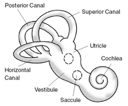
Balance
- The 3 semi-circular canals, along with the utricle and vestibule form the organ that senses our position
 |
| http://en.wikipedia.org/wiki/File:Balance_Disorder_Illustration_A.png |
- The canals are positioned at right angles to each other and movement in any direction will cause the fluid in the corresponding canal to move. Fluid movement in the horizontal canal corresponds to rotation of the head in a horisontal axis, while the posterior and superior canals correspond to vertical movments of the head. Movement of the fluid causes movement of the cilia, which sends electrical impulses to the brain.
- Otoliths are small crystaline structures found in the utricle and vestibule. They rest in a jelly-like matrix over receptors cells. Their movement mechanically stimulates the receptor cells, giving information about gravity and linear motion
 Békésy György (1899-1972), biophysicist, worked in Budapest, Stockholm and the US
-Studied hearing and won a Nobel prize in 1961 for his discovery of the physical mechanism of how stimuation occurs in the cochlea. His research clarified how sound waves travel in the cochlea and how the vibration frequency is associated with stimulation at a specific point.
Bárány Róbert (1876-1936), medical doctor, worked in Austria and Sweden
- Studied the physiology and disease of balance. He developed experimental techniques to study balance and received a Nobel prize for his work in 1914.
No comments:
Post a Comment