Topic 1: The Nervous System

The nervous system is designed to recieve stimuli from inside and outside the body and to respond to them in order to regulate body functions and maintain homeostasis.

The nervous system is built up of specialized cells, which we dealt with when we studied histology (the study of tissues).  You can review it here.  Scroll down to "nervous tissue".

Information is carried through the nervous system along neurons.
Képtalálat

The direction of information flow in a neuron is always from the dendrite to the axon.
Képtalálat

We say that the "nerve impulse" carries the information.  An impulse is actually a change in the cell's charge (an electrical signal).  Neurons have a resting potential of -70mV.  This is the charge of the cell when no information is being sent along it.  When a strong enough impulse arrives, the voltage gated sodium channels found in the cell's membrane open.  This causes the inside of the axon to become more positive than the outside.
Képtalálat
Na-channel opening
The positive charge causes the neighbouring sodium channels to open, allowing the charge change to move along the axon.  Once the initial change in charge has occurred, the sodium channels close and potassium gates open.  Potassium is pumped out of the cell to bring the charge back to -70mV.  The region where this occurs is called the refractory region and a new impulse cannot occur there until it has returned to the resting potential.
Impulse conduction along the axon
At this point, there are more sodium ions inside the cell and more potassium outside the cell.  The sodium-potassium pump plays a role in balancing the difference and also in maintaining a resting potential of -70mV
Képtalálat

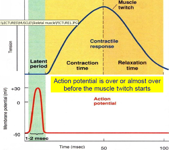
All of what has just been explained is called the ACTION POTENTIAL, which is shown below in its graphical form.
Képtalálat

On the graph, numbers 1 and 4 represent the resting potential, when the cell is not transmitting any information.  When the sodium channels open, the charge of the cell rapidly becomes positive as sodium ions rush in.  This is called depolarization.  Once the sodium channels close and the potassium channels open, the potassium rushes out, causes the charge to become negative again.  This is repolarization.  The return from hyperpolarization (the dip below -70mV) and the maintance of the resting potential is carried out by the sodium-potassium pump.

This video is a nice explanation of the action potential.  

Saltatory conduction refers to the rapid conduction of the impulse down a myelinated axon, where channels opening and closing only occurs in the nodes of Ravier.
Képtalálat

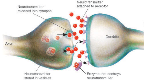
Neurons have spaces between them.  They never actually touch the neighbouring cell.  The axon comes up close to the neighbouring cell's dendrite, but a space remains, which is the synapse.

Képtalálat

Information crosses across the synapse in chemical form.  These chemicals are called neurotransmitters.  The neurotransmitter is released from the axon, it crosses across the cleft (space) and binds to a receptor on the dendrite.
Képtalálat


The attachment to the receptor causes the action potential to begin and pass along the next cell, transmitting the impulse further.

Types of neurons
Neurons can be classified by their function.  Sensory neurons carry signals from the body to the central nervous system (CNS), which is the brain or the spinal cord.  Interneurons pass signals between the sensory neurons and the motor neurons and are found within the CNS.  Motor neurons carry information from the CNS back out to the body in order to "make something happen".

Képtalálat

Neurons are then bundled together to form nerves.  Each neuron is wrapped in a sheath and each bundle of neurons is wrapped in connective tissue.  All the bundles are then held together by more connective tissue to form  nerves made up of hundreds to thousands of neurons.
Képtalálat

Nerves can also be classified.  Motor nerves carry only motor neurons, thus carry information from the CNS to the body.  Sensory nerves are made up of purely sensory neurons, carrying information from the body to the CNS.  Mixed nerves are made up of both motor and sensory neurons, thus carry information in both directions.

THE CENTRAL NERVOUS SYSTEM (CNS)

The CNS is made up of the brain and the spinal cord.  The brain is found in the skull (which provides physical protection) and is surrounded by 3 layers of meninges (pia matter, arachnoid matter and dura matter), which provide further protection.
Képtalálat

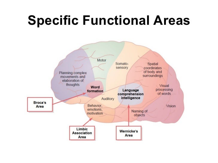
The human brain has multiple areas, the largest of which is the 
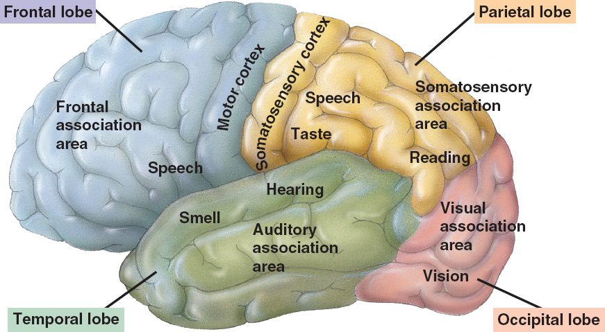
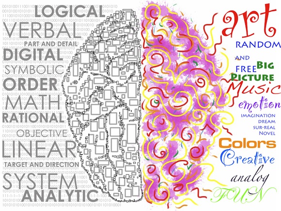
cerebrum (nagyagy).  It is considered to be responsible of the higher functions of the brain.  The cerebrum is divided into two hemispheres.  It was once believed that the left and right sides of the brain took on slightly different roles, with the left brain being associated with logic and language, while the right brain was associated with creativity and being artistic.  Modern research is showing that it is not quite that simple.  Nonetheless, the right brain does control motor function of the left side of the body and the left brain controls motor function on the right side of the body.  The cerebrum is also divided up into lobes.  At the front are the frontal lobes which are associated with reasoning and problem solving and are the last part of the brain to complete development.  Behind the frontal lobes are the 
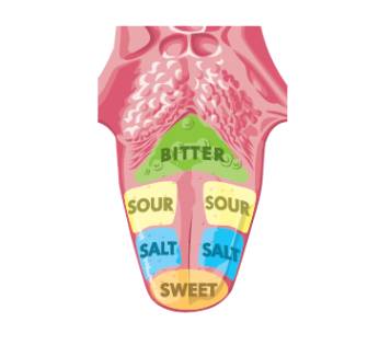
parietal lobes, which are associated with touch, taste, reading and language.  At the very back of the cerebrum are the occipital lobes, which are associated with vision, while at the sides of the cerebrum are the 
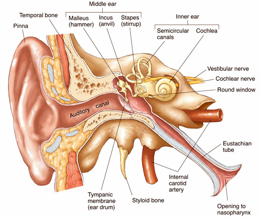
temporal lobes, which are associated with hearing.
Képtalálat
External side-view of the brain.  The wrinkly portion is the cerebrum, the small brownish part is the cerebellum.

Image result
Lobes of the cerebrum


Image result

Below and at the back, the cerebellum (kisagy) is also visible.  It plays an important role in balance and coordination, as well as precision and accurate timing of motions.
Képtalálat
Cross-section of the brain, showing the internal structures
Between the cerebrum and the thalamus is the corpus callosum, which allows the two hemispheres to communicate with each other.

In the middle region of the brain, the thalamus can be found.  It is involved in sensory perception and it controls sleep and awake states of consciousness.  It is thought of as a kind of hub of information, since every sensory system (except smell) travels through the thalamus on its way to the cerebrum.

The hypothalamus and the pituitary gland control the body's hormonal systems.  They both produce hormones and the pituitary stores hormones until they are needed.  These hormones act upon other hormone producing glands, to stimulate hormone production.  The pituitary also produces some hormones that have direct effects on body temperature, production of urine and growth.

The brain stem is composed of the midbrain, the pons and the medulla oblongata.  The midbrain plays a role in body movement, vision and hearing.  The pons relays signals from the forebrain to the cerebellum and affects respiration, swallowing, equilibrium, sleeping states, etc.  The medulla oblongata is responsible for multiple involuntary function, like sneezing, etc.


The spinal cord

Image result

A long bundle of nervous tissue that extends from the brain.  It is part of the central nervous system.  It transmits signals from the brain to the rest of the body.  By itself, it can control numerous reflexes.  It is protected by meninges and cerebrospinal fluid.

Image result
Cross-section of the spinal cord
The spinal nerves enter the spinal cord at its sides.  Sensory information arrives from receptors through the dorsal root ganglion into the grey matter in the middle of the spinal cord.  The sensory neuron will then pass the impulse to an interneuron or directly to a motor neuron.  Motor neurons carry the impulse out of the spinal cord through the ventral root of the nerve.  

Grey matter makes up the central portion of the spinal cord and it is generally cell bodies and dendrites, while white matter make up the dorsal, ventral and lateral parts of the cord and contains the myelinated axons of neurons running up or down the length of the spinal cord.  It is the myelination that gives the white matter its colour.  This area is divided into ascending and descending tracts of neurons.  

Pyramidal and Extrapyramidal Tracts
 
The pyramidal tracts run from the cerebrum through the midbrain and pons to the medulla oblongata, where they cross-over and continue down the spinal cord on the opposite side.  Thus, motor innervation of the right side of the body is controlled by the left side of the brain and vice versa.  The pyramidal tracts control all intentional movement and learned fine motor coordination, like writing.

Image result

The extrapyramidal tracts include parts of the cerebrum, midbrain, cerebellum and brainstem.  These are pathways for coordination of movement and control of muscle tone, as well as controlling large motor movements  and motions that reflect emotions.  The extrapyramidal system is very ancient with 3 of the 4 pathways found in humans being shared with salamanders!!  
Image result


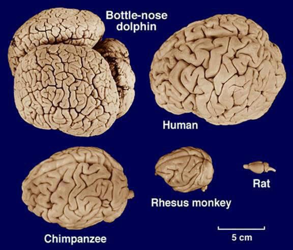
Why is your brain wrinkly?  More wrinkles = larger surface area = more neurons = smarter!  (okay it is a bit more complicated than that, but that is the general idea).
Image result


Functional divisions of the nervous system
Image result

The functionsal divisions of the nervous system refer to what that part of the nervous system does, the situations that it controls.  The somatic nervous system is voluntary control of skeletal muscles, whereas the autonomic system is the involutary control of body functions.
Image result


The details of how the different system function are shown below, if you are really interested. :)

Image result


The parasympathetic system is in charge of involuntary body functions when we are relaxed and resting.  The typical effects are shown below.  The sympathetic system kicks in when we are in a state of fear or great excitement.  It is often referred to as the "fight or flight" response because it is designed for dealing with situations that we should run away from or battle with (eg. sabre-tooth tiger).  Its typical effects are also shown in the image below.
Image result


Motor Function of the Nervous System

Muscle tissue contracts in response to appropriate nervous impulses.
- impulse travels along a motor neuron to the nerve-muscle synapse (neuromuscular junction)
- at the synapse, neurotransmitters (acetylcholine) are released into the synaptic cleft
- neurotransmitters bind to receptors on the muscles, initiating a depolarization process in the muscle, causing actin filaments to pull in between the myosin filaments, resulting in contraction of the muscle
- once the action potential has passed, the muscle will relax
http://www.hoops.co.il/?p=14856
http://www.zoology.ubc.ca/~gardner/chemical_synapses%20-%20postsynaptic.htm


In skeletal muscle:
- one impulse results in one contraction - this is called muscle twitch (izomrángás)

http://howmed.net/physiology/skeletal-muscle/
- since the action potential has passed by the time the muscle contracts, a new action potential can innervate the muscle before the period of relaxation begins.

In the figure below, in (a) we can see a series of twitches, where the impulses are spaced out in time

Staircase effect
- if impulses arrive one after the other in more rapid succession, we can observe what is referred to as the staircase effect, where each twitch is a bit stronger than the one before.  See (b).  Basically, the second twitch came so quickly that the first one never fully relaxed, so there is leftover calcium in the sarcoplasm.  Since there is some force leftover from the first twitch, they build on each other.  This is called summation of force

Tetanus
- In (c), the twitches are created by such a rapid succession of impulses (at least toward the end), that the individual twitches summate and blend into a smooth sustained contraction, called tetanus.  This is also called frequency summation


summation.jpg (15507 bytes)
http://faculty.stcc.edu/AandP/AP/AP1pages/Units5to9/unit9/summatio.htm
- sustained contractions are normal in living organisms, individual twitches are rare.

Muscle tone/Tonus
- skeletal muscles are constantly in a state of passive, partial contraction, which is called muscle tone
- it helps maintain posture and it decreases during REM sleep
- muscles are always ready to respond to sudden changes (eg. in balancing)
- muscle tone is primarily controlled at the level of spinal reflexes, but some aspects also have higher control.
eg. knee-jerk reflex
http://kids.britannica.com/comptons/art-156424/Knee-jerk-reflex-reaction-and-motor-neuron-connection-to-spinal
- hitting the hammer to the knee causes a slight stretch in the quadriceps (thigh muscle on front of leg)
- the stretch receptor in the quadriceps sends an impulse along the sensory neuron to the spinal cord
- the axon branches to two synapses: one to an inhibiting interneuron which synapses with a motor neuron that leads to the biceps muscle and prevents it from contracting; the other branch of the axon synapses directly with a motor neuron which sends and impulse to the quadriceps causing it to contract.
- lower leg kicks forward

- reflexes play a role in more complex motions, but control over most motion occurs at higher levels of the central nervous system
- the organization and coordination of our motions occurs in the cerebrum, in the posterior region of the frontal lobe
-motor tracts descend through one of 2 systems:  pyrmidal and extrapyramidal motor systems

Pyramidal motor system:
- the pyramidal tracts descend from the cerebrum through the brainstem to the grey matter of the spinal cord
- in the brainstem some fibres connect to motor neurons of brain nerves, but the majority cross-over in the pyramids of the medulla oblongata and continue into the spinal cord where they synapse with motor neurons
- a few nerves don't cross-over in the medulla oblongata, but continue straight down into the spinal cord , where they cross-over right before synapsing with motor neurons
- in the end, the motor innervation of the right side of the body is controlled by the left side of the brain and vice versa.
- the pyramidal motor system controls all intentional movement and learned fine motor coordination (like writing)

http://teachmeanatomy.info/neuro/pathways/descending-tracts-motor/


Extrapyramidal motor system:
- includes parts of the cerebrum, the mesencephalon and the cerebellum, as well as the brainstem
- this system controls automatic learned movements, large motor movements and motions that reflect emotions, as well as taking part in maintaining muscle tone
- the extrapyramidal system is very ancient and 3 of the 4 tracts found in humans are shared by salamanders

-well-controlled movement requires the coordinated function of both the pyramidal and the extrapyramidal systems

- the contraction of smooth muscle is also controlled by stimulation from vegetative motor neurons.
- contraction of the heart muscle does NOT require external stimulation.  The heart contains small muscle cells that are capable of spontaneous stimulation.  These cells are more permeable to sodium than the others and are found grouped at the sino-atrial node and at the atrio-ventricular node.


Sensory function of the nervous system

- information from the internal and external environments  is collected by receptors and brought to the central nervous system through spinal and cerebral nerves.
- stimulation of receptors in skin and muscle is transferred to spinal sensory nerves (found in the dorsal ganglion), which carry the stimlation through the sensory tracts of the spinal cord
- some take part in spinal reflexes, these pass into the ventral horn and synapse with motor neurons there.
- most continue on the ascending tracts and cross-over in the spinal cord or the medulla oblongata and enter cerebral centers on the opposite sides.

- stimulation from the touch and heat receptors of the skin enters the dorsal horn, then cross-over in the spine before continuing along the lateral ascending tracts to the thalamus.
- sensory information from the deeper layers of the skin and the muscles enters the dorsal ascending tracts to the medulla oblongata, there the neurons synapse, then cross-over and travel along the opposite side to the thalamus
- information is processed and grouped in the thalamus, then sent on to the appropriate parts of the cerebrum.
http://faculty.southwest.tn.edu/rburkett/A&P1_nervous_syst_organization.htm
Taste receptors are found in tastebuds on the tongue.  The stimulation is carried by cranial nerves to the medulla oblongata and from there to the thalamus and the cerebrum.  All flavours are derived from sweet, salty, sour and bitter.  Typically, it is stated that sweet receptors are at the tip of the tongue, bitter is at the back and the sides contain salty and sour.  I have recently read that this is not entirely true, but....  that is what is in the textbook :)

http://chargedmagazine.org/2012/09/taste-buds/
Smell receptors or olfactory receptors are found on the back surface of nasal passage
http://www.allthingsherbal.biz/2011/10/aromas-and-the-sense-of-smell/

- stimulation of the cerebral olfactory nerves goes directly to the cerebrum without passing through the thalamus

Sight receptors are the eyes! They contain many light receptors, but the eye is more complex that simply being a mass of light receptors.

The eyeball is surrounded by 3 layers:
- the outer layer is protective, the sclera (ínhártya) is is the white layer, which becomes the clear cornea (szarúhártya) at the front of the eye.  Light passes through the cornea to the inside of the eye.  The cornea is constantly moistened by the blinking of the eyelids.
- the middle layer contains many blood capillaries, providing the eye with necessary oxygen and nutrients, it also absorbs excess light to prevent damage.  It is called the choroid (érhártya).  At the front it forms the ciliary body, which to which the muscles controlling the lens attach.  The iris (szivárványhártya) is a continuation of the ciliary body and the hole in the middle, through which light passes into the eye is called the pupil.  The colour of the iris depends on the number of pigments it contains.  If someone's eyes are black or brown, then the iris contains many pigments, while people with green or blue eyes contain fewer pigments.  
- the inner layer is where the receptors are found.  It is called the retina (ideghártya) and it contains two kinds of light receptors, rods and cones.

http://libot-libot.blogspot.hu/2011/05/cataract-is-clouding-of-lens-of-eye.html

http://lpatersonbiotask3.wikispaces.com/Describe+the+anatomy+and+function+of+the+human+eye...

- light passes through several different media before it reaches the receptors
- First the light passes through the cornea
- Then it passes through space between the cornea and the lens, which contains the aqueous humour (csarknovíz), which is watery in consistency
- The size of the pupil decreases when there is lots of light, due to the constriction of the iris, and increases in low light, due to relaxation of the iris.  
- Light then passes through the lens (just behind the iris).  The lens is flexible and can be flattened or rounded to focus the light on the fovea (sárgafolt) at the back of the retina.  This is the point in the eye of truly focused vision
-  When the eye is relaxed then focus is on distant objects and the muscles holding the lens are constricted, so the lens is flattened, the opposite occurs when focusing on close objects, the muscles relaxes and the lens becomes rounded. With age, the flexiblilty of the lens decreases, so most elderly people have good distant vision, but difficulty focusing close objects.  Focal problems can and often do occur earlier in life and can be corrected with proper optical lenses.  If someone is far-sighted the focal point is behind the retina, while if someone is near-sighted the focla point is in front of the retina.
- the chamber behind the lens is filled with the vitreous humour, which is a jelly-like substance that helps maintain the eye's shape. 
- when light reaches the retina, it activates the receptors, the rods (pálcikák) and cones (csapok), which got their names from their unique shapes.
- Rods are long and cylindrical.  They work well in low light and produce black and white images.
- Cones are smaller and conical.  They function well in bright light and allow us to see in colour.
- When photons are "caught" by the receptors, their energy is transformed into an electrical impulse, which is transfered down the receptor cell's membrane to the synapse.  The stimulation is then carried by sensory neurons in the optic nerve to the thalamus.  From here, via synapses, the stimulation is sent to the visual areas in the occipital lobes.

http://www.biochemj.org/csb/010/csb010_fig071.htm
Rods and cones are organized as shown below.  Where the optic nerve enters the back of the eye, there is a point on the retina where there are no rods or cones.  This is the blind spot.
http://tiffanybiology.blogspot.hu/2011/05/rods-and-cones.html


http://abcarcade.com/blindspot-test.html


Sound and balance receptors are found in the ears.

http://www.biographixmedia.com/human/ear-anatomy.html
Sound
- The outer ear's shape is designed to bring sounds into the auditory canal to the ear drum (also called tympanic membrane).
- The ear drum is the beginning of the middle ear.  In the space behind the ear drum, the smallest bones in the body called ossicles, the hammer, anvil and stirrup, can be found.  The hammer moves when a sound vibration causes the ear drum to vibrate.  The movement of the hammer moves the anvil, which in turn, moves the stirrup.  The bottom of the stirrup hits the oval window (not labelled in upper diagram) of the inner ear.
- The inner ear begins with the oval window and beyond it, the cochlea, which is filled with fluid called perilymph.  When the stirrup hits the oval window, this begins a wave in the cochlear fluid.  The round window, at the other end, gives the fluid somewhere to go.
- The basilar membrane is a rigid surface that extends the length of the cochlea.  When the stirrup moves in and out, it pushes and pulls on the basilar membrane that is right below the oval window.  This force starts a wave along the surface of the membrane.  The basilar membrane has a peculiar structure.  It is made of about 25 000 reed-like fibers that extend the width of the cochlea.  Near the oval window the fibers are short and stiff.  Toward the other end the fibers are longer and more flexible.  Thus the fibers have different resonant frequencies.  A specific wave frequency will resonate perfectly with the fibers at a certain point, causing them to vibrate rapidly.  When the wave reaches the fibers with the same resonant frequency, the wave's energy is suddenly released.  Because of the increasing length and decreasing rigidity of the fibers, higher-frequency waves vibrate close to the oval window and lower frequency waves vibrate at the other end of the membrane.


http://bio1152.nicerweb.com/Locked/media/ch49/SAVE/cochlea.html

-  The organ of Corti is a structure containing thousands of tiny hair cells.  It lies on the surface of the basilar membrane.  When a wave finally reaches the resonant point, the membrane suddenly releases a burst of energy in that area.  This energy is strong enough to move the organ of Corti hair cells at that point.  When the hair cells move, they send electical impulses through the cochlear nerve to the thalamus and then to the cerebral cortex, where the brain interprets them.  Louder sounds release more energy at the resonant point, thus move greater numbers of hair cells in that area.

http://bio1152.nicerweb.com/Locked/media/ch50/cochlea.html

http://nl.bu.edu/research/projects/moneta/moneta-v2-0/auditory-system/

 Balance
- The 3 semi-circular canals, along with the utricle and vestibule form the organ that senses our position
File:Balance Disorder Illustration A.png
http://en.wikipedia.org/wiki/File:Balance_Disorder_Illustration_A.png
- The semi-circular canals are filled with a fluid called endolymph and lined with tiny cilia
- The canals are positioned at right angles to each other and movement in any direction will cause the fluid in the corresponding canal to move.  Fluid movement in the horizontal canal corresponds to rotation of the head in a horisontal axis, while the posterior and superior canals correspond to vertical movments of  the head.  Movement of the fluid causes movement of the cilia, which sends electrical impulses to the brain.
- Otoliths are small crystaline structures found in the utricle and vestibule.  They rest in a jelly-like matrix over receptors cells.  Their movement mechanically stimulates the receptor cells, giving information about gravity and linear motion    

Békésy György (1899-1972), biophysicist, worked in Budapest, Stockholm and the US
-Studied hearing and won a Nobel prize in 1961 for his discovery of the physical mechanism of how stimuation occurs in the cochlea.  His research clarified how sound waves travel in the cochlea and how the vibration frequency is associated with stimulation at a specific point.

Bárány Róbert (1876-1936), medical doctor, worked in Austria and Sweden
- Studied the physiology and disease of balance.  He developed experimental techniques to study balance and received a Nobel prize for his work in 1914.


Nervous Control of Human Behaviour

Almost all information about our environment that is collected by receptors is taken to the cerebral cortex.
The cortex's massive neural network processes, stores and, if necessary, recalls the information.  This is where sensory information becomes emotions, where responses are organized and impulses are sent out through motor neurons.

The Limbic System
The limbic system is a complex set of brain structures that lie on both sides of the thalamus, right under the cerebrum.  It is not a separate system, but a collection of structures including olfactory bulbs, hippocampus, amygdala and hypothalamus.
http://webspace.ship.edu/cgboer/limbicsystem.html
It supports a variety of functions including emotion, behaviour, long-term memory, motivation and olfaction (smelling).  It can be divided into 2 functional areas, the inner and outer rings.  The outer ring is closely associated with the hypothalamus, thus in this way the limbic system is in direct connection with the hormonal system.  The other main function of the outer ring is the control of emotional behaviour, including such emotional reactions like pain, joy, fear  and anger.  These emotions are associated with specific areas in the outer ring.    The inner ring is primarily associated with the storage of important memories.  The memories are not actually stored in the limbic system, but it controls where memories will be stored in the cerebrum.  Memories can be short-term memories, which only last for a few minutes before being replaced by other information, or they can be long-term memories which are stored for many years, if not whole lifetimes.  The long-term storage of memories requires the creation of synapses. The more often a synapses is activated the greater the amount of neurotransmitter, thus creating a more stable synapse and a more lasting memory.

Memory
Memory and memory storage is an area of constant research.  To read a good, clear overview of memory creation, storage and retrieval click here.

Features of human memory:
1. Memorization is easier if the information as meaning to the individual.
2. Well-organized information is easier to retrieve.
3.  Storage and retrieval of information is easier if them memory can be associated with other key stimuli.
4.  Storage is improved if there is emotional weight to the memory.
5. Interference between memories makes storage and retrieval more difficult (eg. learning large amounts of new material can make it more difficult to retrieve old information)

Speech
Speech assists humans in their social interactions.  At birth humans are capable of creating nearly 200 different sounds (this is a biological ability).  In any given social environment (nation, culture), only 25-45 of these sounds are used.   Humans also use writing and gestures (metacommunication) to communicate.  In greater than 85% of people the speech association areas are found in the left lobe of the cerebrum.  The auditory association area, found in the temporal lobe, also takes part in the creation and understanding of speech.  Most of the time, it is also in the left brain that these sounds are converted into thoughts.  


The Cerebral Cortex
The cerebral cortex, often referred to as grey matter, is considered to be responsible for "higher" brain function.  The surface of the cortex is folded in large mammals, increasing its surface area.  
http://gerardkeegan.com/glossary/cerebral-cortex
The two halves of the cerebrum are asymmetreical, both in structure and in function.  The two sides complement each other: while one side takes part in speech and deduction, the other deals with physical and temporal connections (representations).  In women, this asymmetry is less apparent.
http://www.as.wvu.edu/~rbrundage/chapter5b/sld014.htm
http://bio1152.nicerweb.com/Locked/media/ch48/cerebral.html
Asymmetry of the cerebrum